

A Comprehensive Selection
for Veterinary Care
Samsung ultrasound systems for veterinary adopt Samsung’s pioneering
imaging engine, Crystal Architecture™, boast various advanced features,
easy-to-use operations, and dedicated design that improves the
routine diagnostic experience.
Redefined imaging technologies powered by Crystal Architecture™
.png)
Crystal Architecture™, an imaging architecture that combines CrystalBeam™ and CrystalLive™, based upon S-Vue Transducer™, provides a crystal clear image. CrystalBeam™ is a new beamforming technology beneficial in delivering high-quality image resolution and increased uniformity of images. CrystalLive™ is Samsung’s up-todate ultrasound imaging engine with enhanced 2D image processing,3D rendering and color signal processing, to offer outstanding image performance and efficient workflow during complex cases.
Sophisticated 2D Image Processing
Provide uniform imaging
performance of overall image area
S-Harmonic™mitigates the signal noise, enhances contrast, and provides uniform image performance of overall image area from near-to-far.

Abdomen

Abdomen with S-Harmonic™ ᵃ
Reduce noise to improve 2D image quality
ClearVision,is a noise reduction filter that enhances the edge contrast and creates sharp 2D images for optimal diagnostic performance. In addition, it provides application-specific optimization and advanced temporal resolution in live scan mode.

Kidney

Kidney with ClearVision
Enhance hidden structures in shadowed regions
ShadowHDR™ selectively applies high-frequency and low-frequency of the ultrasound to identify shadow areas where attenuation occurs.

Intercostal view

Intercostal view
with ShadowHDR™ ⁴
Clean up blurry areas in the image
HQ-Vision™provides clearer images by mitigating the characteristics of ultrasound images that are slightly blurred than the actual vision.

Quadriceps tendon

Quadriceps tendon
with HQ-Vision™ ⁴
Spatial and contrast resolution with artifact suppression
MultiVisioncontrols ultrasound beam electronically by steering, and compounds multiple scan lines for better image. MultiVision provides remarkable spatial and contrast resolution with even greater artifact suppression than ever before.
Detailed Expression of Blood Flow Dynamics
Provide uniform imaging
performance of overall image area
S-Harmonic™mitigates the signal noise, enhances contrast, and provides uniform image performance of overall image area from near-to-far.

Kidney S-Flow™ with LumiFlow™ ᵃ
Visualize slow flow
in microvascular structures
MV-Flow™ ¹visualizes microcirculatory and slow blood flow to display the intensity of blood flow in color.

Kidney with ClearVision
Show blood flow in vessels
in a 3D-like display
LumiFlow™ a function that visualizes blood flow in three dimensional-like to help understand the structure of blood flow and small vessels intuitively.

Kidney MV-Flow™ with LumiFlow™ ᵃ
Contrast Enhanced Ultrasound
CEUS+ ¹ is a contrast agent imaging technology. The micro-bubble contrast agent injected into the body through the vein or alike is subjected to perform nonlinear resonance due to stimulation of ultrasound energy.
.png)
Kidney S-Flow™ with LumiFlow™ ᵃ
Enriched Diagnostic Features
Hepato-renal index with
automated ROI recommendation
HRI (Hepato Renal Index)is an index to quantify steatosis of a liver by comparing echogenicity between liver parenchyma with renal cortex. EzHRI™ places 2 ROIs on the liver parenchyma and renal cortex and provides HRI ratio.

Kidney S-Flow™ with LumiFlow™ ᵃ
Quantitative measurement of
tissue attenuation
TAI™ ¹ provides quantitative tissue attenuation measurement to assess steatotic liver changes.

Kidney with ClearVision
Quantitative measurement of
scatter distribution
TSI™provides quantitative tissue scatter distribution measurement to assess steatotic liver changes.

Kidney MV-Flow™ with LumiFlow™ ᵃ
Display and quantify tissue stiffness
in a non-invasive method
S-Shearwave Imaging™ allows for non-invasive assessment of stiff tissues in various applications. The color-coded elastogram, quantitative measurements, display options, and user-selectable ROI functions are useful for accurate diagnosis of various regions

Kidney S-Flow™ with LumiFlow™ ᵃ
Measure Ejection Fraction of the
left ventricle conveniently
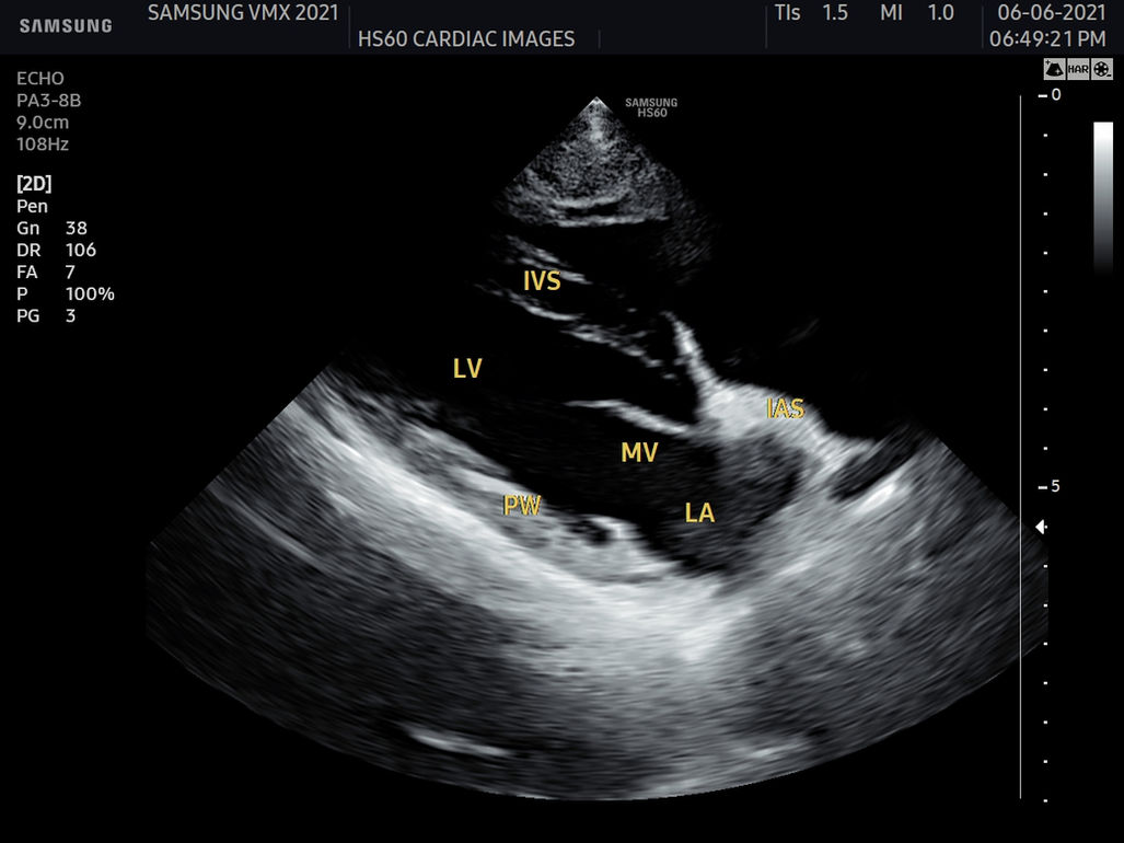
AutoEF is a feature which measures and quantifies Ejection Fraction. By selecting the three points of the left ventricle, the volume at the end-systolic and end-diastolic points of the left ventricle is calculated, to assist in quick and efficient assessment.

Kidney MV-Flow™ with LumiFlow™ ᵃ
Display in extended field-of-view
Panoramic+imaging displays as an extended field-of-view so users can examine wide areas that do not fit into one image as a single image. Panoramic+ imaging also supports angular scanning from linear transducer data acquisition.

Panoramic+ ⁴
Display needle tip clearly
NeedleMate+™ ¹delineates needle location when performing interventions such as nerve blocks. Improved accuracy and efficiency in procedure are possible with beam steering added to NeedleMate+™.

NeedleMate+™ ⁴


Improved Workflow Efficiency
and Ergonomic Design
We believe that a truly great system offers customer-centric working conditions. The streamlined workflow
supports your daily procedures by reducing keystrokes and by combining multiple actions into one. Users
have the option of customizing their diagnostic settings based on personalized protocol, resulting in a more
simplified exam process and faster workflow.
Optimize image with
one touch of the button
QuickScan™ technology provides intuitive optimization of both grayscale and Doppler parameters.
Build predefined protocols to ensure every step is followed every time
EzExam+™ ensures the full investigation is performed, eliminating the risk of forgetting an image or loop capture, as well as measurement and transducer preset changes.
Compare previous and current exam
in a side-by-side display
EzCompare™ automatically matches the image settings, annotations, and bodymarkers from the prior study.
Select transducer and preset
combinations in one click
QuickPreset allows the user to select the most common transducer and preset combinations in one click.
Customize frequently used functions on the touchscreen
A customizable touchscreen allows the user to move frequently used functions to the first page.
Tilt touchscreen to accommodate user preference
Samsung’s tilting touch screen can be adjusted to accommodate user’s viewing preferences in any scanning environment.
Use the system when AC power is temporarily unavailable
BatteryAssist™ provides battery power to the system, enabling users to perform scans when AC power is temporarily unavailable.
Save image data directly to
USB memory
QuickSaveA customizable touchscreen allows the user to move frequently used functions to the first page.
Reference tool to practice the
procedures during live scanning
As a reference tool for plane scanning, EzAssist™ has multiple clinical applications during live scanning. This allows clinicians to practice procedures using valuable reference materials such as clinical reference images and animation.
Real-time image streaming solution
SonoSync™ ³ is available in PC and smartphone, etc. as a real-time image share solution that allows communication for care guide and training between doctors and sonographers.
In addition, voice chatting, text chatting and real-time marking functions are provided for better communication; and the MultiVue function is included that allows monitoring multiple ultrasound images on a single screen.

Sleep Mode
It reduces boot-up time by using sleep mode without having to shut down or restart the system.
Diverse Clinical Cases and Veterinary Types
Related Products
RS85 Prestige
The Real Revolution
RS85 Prestige has been revolutionized with novel diagnostic features across each application based on the preeminent imaging performance. The advanced intellectual technologies are to help you confirm withconfidence for challenging cases, while the easy-to-use system supports your effort involved in the routine scanning.
Sophisticated 2D Image Processing
ShadowHDR™, HQ-Vision™, PureVision™, S-Harmonic™, ClearVision, MultiVision
Detailed Expression of Blood Flow Dynamics
MV-Flow™ ¹, S-Flow™, LumiFlow™ ¹ , CEUS+¹
Enriched Diagnostic Features & Interventional Solutions
ElastoScan™ ¹, ², S-Shearwave™ ¹, S-Shearwave Imaging™ ¹, Strain+¹, Panoramic+, NeedleMate™
Enhanced Productivity and Facilitated Workflow
EzExam+™, EzPrep™, QuickScan™, QuickPreset, Touch Customization, Sonosync™ ³
Ergodynamics for Your Comfort
6 Way Control Panel, Central Lock, Maneuverable Wheel, Gel Warmer, 23.8-inch LCD Monitor, 14-inch Tilting Touch Screen

Comprehensive selection of transducers
Curved array transducers


CA1-7S*
CA1-7S*
CA3-10A
CA2-8A
CA4-10M*
Linear array transducers


LM2-18
LA2-14A
LA2-9S*
LA2-9A
LA3-16A
LA4-18A*
LM4-15B
LA3-22AI
Phased array transducers


PA1-5A*
PA3-8B
PA4-12B
* Ergonomic transducers
These transducers have a newly designed ergonomic hand-grip and better weight distribution for comfortable scanning
Secure Your Care
Samsung Healthcare Cybersecurity
Bringing peace of mind to your hospital and patients
To address the emerging need for cybersecurity, Samsung provides a solution to support our customers by offering the tools to protect against cyberthreats that may compromise invaluable patient data and ultimately degrade the quality of care. Samsung’s Cybersecurity Solution strives to abide by the CIA triad (Confidentiality, Integrity, and Availability) and takes a comprehensive approach to providing impeccable protection with the following pillars: Intrusion prevention, Access control, and Data protection
Intrusion Prevention
Tools for protecting against cyber
threats from external attacks
-
Security tools include
Anti-virus & Firewall
-
Secured operating system
Access Control
Strengthened surveillance for tracking
the access of patient information
Account management
Enhanced audit trail
Intrusion Prevention
Encryption functions for safeguarding
data whether at-rest or in-transit
-
Data protection
-
Transmission security
-
The products, features, options, and transducers may not be commercially available in some countries.
-
Sales and Shipments are effective only after the approval by the regulatory affairs. Please contact your local sales representative for further details.
-
This product is a medical device, please read the user manual carefully before use.
-
Prestige is not a product name but is a marketing terminology.
1. Optional feature which may require additional purchase.
2. Strain value for ElastoScan+™ is not applicable in Canada and the United States.
3. SonoSync™ is an image sharing solution.
4. This is not an image of the animals, but just provided to help you better understand the feature.
a. Image acquired by RS85 Prestige ultrasound system.
b. Image acquired by V8 ultrasound system.
c. Image acquired by HS60 ultrasound system.
d. Image acquired by HS40 ultrasound system


.jpg)


.jpg)


.jpg)


.jpg)


.jpg)


