


All the key benefits
you want
The V7 offers a fascinating performance and gives you the possibility to do
what you want with comprehensive tools that feature the latest
innovations.
For instance, ViewAssist™'s amazing features automatically perform
measurements and annotations with a simple click of a button, thereby
reducing repetitive tasks for healthcare professionals.
Rich in features, V7 is fully capable of covering women's health that
allows you to explore to the fullest.
Diagnose diverse and challenging clinical cases
The V7 comes with a variety of tools for diverse and challenging cases.
Healthcare professionals can execute targeted examinations with ease, using the necessary advanced features prepared in the right place.
Furthermore, various sophisticated 2D and color imaging features are supported for extraordinary image quality.
2D imaging


3D imaging


Color imaging


Diagnostic features


Enriched diagnostic features
with accuracy and precision
The V7 system comes with advanced features that assist in precise diagnosis and increasing throughput.
The V7’s variety of features and user-friendly interface aid in significantly improving the healthcare professionals’ daily ultrasound examination experience.


Measure the size and shape of
the uterus with AI technology
UterineAssist™ ¹, based on Deep Learning technology, automatically measures the size and shape of the uterus, assisting in detecting signs of uterine-related abnormalities, as well as reducing scan time.


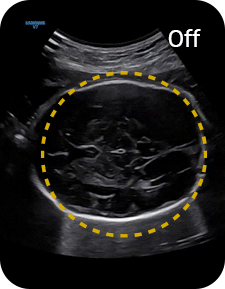
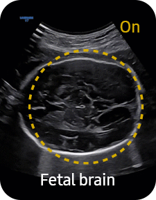
An automated classification and
annotation of the images
ViewAssist™ ¹ provides automatic classification of the ultrasound images and annotation of the structures to help healthcare professionals in convenient measurement.

Uterine Contour
Uterine Contour automatically extracts the centerline and thickness of the curved endometrium and provides a coronal view in 3D, flattened by the centerline. In addition, uterine malformation classification are reported according to the ESHRE/ESGE* or ASRM** guideline selection.time.
* ESHRE/ESGE : The European Society of Human Reproduction and Embryology / The European Society for Gynaecological Endoscopy
** ASRM : The American Society for Reproductive Medicine
Support in deciding
delivery method
LaborAssist™ ¹ provides information about the progress of delivery from the automatic measurement of the AoP (Angle of Progress) and the direction of the fetal head. This helps in making delivery decisions and effective communication with the mother about the delivery process.
* ASRM : The American Society for Reproductive Medicine
Examine patency of the fallopian tube and
morphology of uterus and endometrium
CEUS+ HyCoSy ¹can be used in 3D/4D for effective examination for patency of the fallopian tube and morphology of uterus and endometrium. 4D Prospective storage allows 4D data to be stored at the same time the contrast agent is injected.
Examine patency of the fallopian tube and
morphology of uterus and endometrium
5D Heart Color™ ¹identifies 9 standard planes of the heart using fetal STIC data and important information about fetal heart development, complying with AIUM guidelines. It also offers dedicated Preset, Predictive Cursor, Diagnostic Alert, and heart Diastole/Systole time points.
Measure stiffness of cervix area
for predicting preterm birth
E-Cervix™ ¹ measures the stiffness of the cervical area. Using elasticity images that help predict preterm birth and induced labor, it enhances reproducibility and reduces inter observer variation by using a sum of various elastograms acquired for several seconds.
Analyze selected thyroid lesions
and report thyroid assessment
S-Detect™ for Thyroid ¹, ⁴ analyzes selected lesions in the thyroid ultrasound study and shows the analysis data, provides standardized reporting based on the ATA, BTA, EU-TIRADS, and K-TIRADS* guidelines; and helps diagnosis with the streamlined workflow.
*ATA: American Thyroid Association
BTA: British Thyroid Association
EU-TIRADS: European Thyroid Imaging Reporting and Data System
K-TIRADS: Korean Thyroid Imaging Reporting and Data System
Estimate fetal weight to check the
growth of the fetus
5D Limb Vol.™ is a semi-automated tool to quickly and accurately measure upper arm or thigh volumes from 3 simple seed points on a single volume data set. These measurements can then be used to calculate an accurate estimation of fetal weight.

Analyze selected breast lesions
and report breast assessment
S-Detect™ for Breast ¹, ⁴analyzes selected lesions in the breast ultrasound study and shows the analysis data, applies BI-RADS ATLAS* to provide standardized reporting; and helps diagnosis with the streamlined workflow.

* Breast Imaging-Reporting and Data System, Atlas
It is a registered trademark of ACR and all rights reserved by ACR.
Display tissue stiffness in color image
A diagnostic ultrasound technique for imaging elasticity, ElastoScan+™ observes the transformation of the tissue strain by the internal or external forces, and converts relative stiffness into a color image.
Easily calculate the strain ratio between two ROIs
E-Strain™is designed to enable quick and easy calculation of the strain ratio between two regions of interest for day-to-day practice. Simply by setting the two targets, you can receive accurate, consistent results and make informed decisions in many types of diagnostic procedures.
Measure LV MPI and RV MPI
semi-automatically
MPI+is able to semi-automatically measure LV MPI and RV MPI, providing a high reproducibility. After acquiring Inflow/Outflow doppler, RV MPI proceeds alignment by utilizing synchronized signals of the heartrate and valve movement. Through the automatic alignment, it provides ICT, IRT, and RV MPI test results.
Measure the size of follicles
based on 2D imaging
2D Follicle™ ¹ identifies and measures the size of follicles based on a 2D image and provides information about the status during gynecology examinations.
Assess the risk of infertility
using volume data
5D Follicle™ ¹ identifies and measures multiple ovarian follicles in one scan for rapid assessment of follicular size and status during controlled ovarian stimulation.

Extraordinary image quality delivers
diagnostic confidence

Gain insight into complex issues with exceptional image quality and resolution by Samsung’s core imaging engine, Crystal Architecture™.
The proprietary technology combines enhanced 2D image processing and detailed color signal processing to optimize and refine the image.
The cutting-edge V7 will provide outstanding image clarity for a confident diagnosis.


Clean up blurry areas
in the image
HQ-Vision™ ¹ provides clearer images by mitigating the characteristics of ultrasound images that are slightly blurred than the actual vision.


Visualize slow flow in microvascular structures
MV-Flow™ ¹visualizes microcirculatory and slow blood flow to display the intensity of blood flow in color.

Show blood flow in vessels
in a 3D like display
LumiFlow™ ¹is a function that visualizes Blood flow in 3 dimensional-like to help Understand the structure of blood flow and small vessels intuitively.
Efficient workflow
re-designed for simplicity
Made to maximize efficiency, allow V7 to streamline your workflow and reduce various tasks to just a few steps or keystrokes.
The user experience is enhanced through how V7 displays scan data more easily and accurately.

Real-time image sharing, discussion,
and remote control of ultrasound system
SonoSync™ ¹, ⁶ is available in PC and smartphone, etc. as a real-time image share solution that allows communication for care guide and training between doctors and sonographers.
In addition, voice chatting, text chatting and real-time marking functions are provided for better communication; and the MultiVue function is included that allows monitoring multiple ultrasound images on a single screen.
* SonoSync™ is an image sharing solution, not a diagnostic solution.

See images in expanded view
The ultrasound examination can be performed while viewing the images and cines that are expanded at
various ratios according to the user preference.

Simple transfer of fetal ultrasound images and clips
HelloMom™ ¹, ⁵ is a simple and secure image-sharing solution that generates a QR code for the selected fetal images to be transferred. HelloMom™ allows pregnant women and their family to download fetal ultrasound images simply by scanning the QR code with their smartphones, reducing the hassle of installing a separate application.

Easily manipulate volume data
from the touchscreen
TouchGestureintuitively allows you to rotate, zoom, crop, and move 3D images right from the touchscreen.

Customize frequently used
functions on the touchscreen
TouchEdit, a customizable touchscreen, allows the user to move frequently used functions to the first page.
Secure Your Care
Samsung Healthcare Cybersecurity
Bringing peace of mind to your hospital and patients
To address the emerging need for cybersecurity, Samsung provides a solution to support our customers by offering the tools to protect against cyberthreats that may compromise invaluable patient data and ultimately degrade the quality of care. Samsung’s Cybersecurity Solution strives to abide by the CIA triad (Confidentiality, Integrity, and Availability) and takes a comprehensive approach to providing impeccable protection with the following pillars: Intrusion prevention, Access control, and Data protection.
Intrusion Prevention
Tools for protecting against cyber
threats from external attacks
-
Security tools includeAnti-virus & Firewall
-
Secured operating system
Access Control
Strengthened surveillance for tracking
the access of patient information
-
Account management
-
Enhanced audit trail
Intrusion Prevention
Encryption functions for safeguarding
data whether at-rest or in-transit
-
Data protection
-
Transmission security

-
This product, features, options, and transducers are not commercially available in all countries.
-
Sales and Shipments are effective only after the approval by the regulatory affairs.
-
Please contact your local sales representative for further details.
-
This product is a medical device, please read the user manual carefully before use.
-
Optional feature which may require additional purchase.
-
S-Vue Transducer™ is the name of Samsung’s advanced transducer technology.
-
Strain value for ElastoScan+™ is not applicable in Canada and the United States.
-
Recommendations about whether results are benign or malignant in S-Detect™ are not applicable in the United States.
-
A purchase of Mobile Export option is required to use HelloMom™.
-
SonoSync™ is an image sharing solution.
The described product information, including features and options, is CE marked. Regulatory approval/clearance status may vary by country.