

HERA W10
HERA W10, the premier model of the HERA platform, will help you get more insight in diagnosis with its intuitive visualizations,precise analytic features, and instant operation. It is our commitment for Obstetrics and Gynecology applications to supportfor life-long healthcare of women, diligently pursuing for new possibilities in ultrasound diagnosis.
Highlights

Clinical Images


Redefined imaging technologies
powered by Crystal Architecture™
Crystal Architecture™, an imaging architecture that combines CrystalBeam™ and CrystalPure™,
while based upon S-Vue Transducer™, is to provide crystal clear image.
- CrystalBeam™ is a new beamforming technology beneficial in delivering high-quality image resolution and increased uniformity of images.
- CrystalPure™ is Samsung's up-to-date ultrasound imaging engine with enhanced 2D image processing, color signal processing, and advanced intellectual algorithm to offer outstanding image performance and efficient workflow during complex cases.


Fast Rendering
X3 GPU Memory*

Processing Power*
for high-quality images

Data Transfer Rate*
for fast frame rates

* Compared to the Samsung WS80A
A new beamforming for
in-depth image creation
CrystalBeam™ utilizes Arbitrary Waveform Transmission, Massive Parallel Beamforming,and Synthetic Aperture technologies to produce a faster frame rate and improved image uniformity.

Conventional
Arbitrary Waveform
Transmission

Spatial Resolution
Massive Parallel
Beamforming

Frame Rate
Synthetic
Aperture

Frame Rate
* Compared to the Samsung WS80A
Sophisticated 2D & Color Images Processed by CrystalPure™
CrystalLive™ helps you to make more confident diagnoses with fundamental 2D images.Some major advantages of 2D images include shadow-suppressed images, lessened halo artifacts, and mitigated blurred area.
Visualize attenuated shadow area

Intra Uterine Device with CrystalVue™
CrystalVue™ is an advanced volume rendering technology that enhances visualization of both internal and external structures in a single rendered image using a combination of intensity, gradient and position.


Realistic description of 3D/4D performance
CrystalLive™ in 3D/4D provides users with more realistic and high-resolution images.It outdoes conventional 3D imaging technologies in terms of viewing small parts and lighting effects.
Visualization of internal and external structures with volume rendering ¹

Intra Uterine Device with CrystalVue™
CrystalVue™ is an advanced volume rendering technology that enhances visualization of both internal and external structures in a single rendered image using a combination of intensity, gradient and position.


Detailed expression of blood flow dynamics
Color performance of CrystalLive™ has been improved to clearly visualize the hemodynamics of the blood flow.Greater sensitivity resulting from new color signal processing allows for a more accurate detection of peripheral blood vessels,microcirculatory blood flows, and volumes of slow blood flows.
Three dimensional-like visualization of blood flow

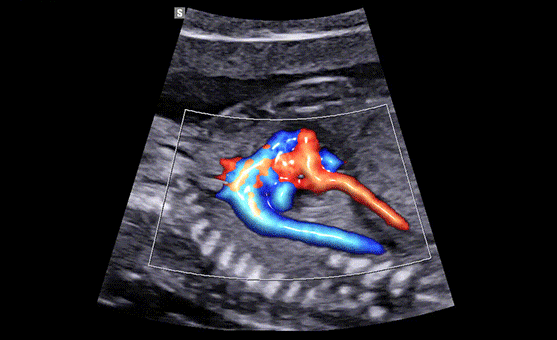
Color Doppler with LumiFlow™ (4 Chamber view)
LumiFlow™ is a three-dimensional visualization of blood flow, which helps to understand the structure of blood flow and small vessels intuitively

Enriched diagnostic system,
excellence in utilization
Images created by the Crystal Architecture™ technologies enhance various diagnostic features of Samsung ultrasound. HERA W10’s diverse technologies to examine the growth of fetus and women’s health in detailed reports will help you build more confidence and enhance the workflow in your diagnosis.

HeartAssist™ ¹
A semi-automated reporting tool for fetal heart diagnosis

Color Doppler with LumiFlow™ (4 Chamber view)
HeartAssist™, while based on big data, it semi-automatically classifies ultrasound image into measurement views required for fetal heart diagnosis and provides measurement results and distribution graph.

ViewAssist™ ¹
Uterine Contour

BiometryAssist™ ¹
A semi-automated reporting tool for fetal heart diagnosis

A semi-automatic technology for biometric measurement, BiometryAssist™, enables users to measure the growth of the fetus quickly while maintaining exam consistency.

Slice A ¹
5D CNS+™ ¹ (Central Nervous System)

5D Limb Vol.™ ¹
Fast fetal weight estimation tool for checking growth of the fetus

5D Limb Vol.™ is a semi-automated tool to quickly and accurately measure upper arm or thigh volumes from 3 simple seed points on a single volume data set.

MPI+ ¹ (Myocardial Performance Index)
FreeForm™
Ergodynamics that Relieves Inconvenience
FreeForm™ refers to Samsung’s new design theme. It was developed to provide a more comfortable diagnostic experienceby reducing the need for movement from one spot to another. Our goal is to satisfy user’s working environment by applying a mechanismto the control panel in its widemoving range, as well as by considering a user’s arm reach. This enables it to offer a sufficientamount of space for the user’s knee.


Control Panel Moving Mechanism
An internal study showed that Samsung’s Control Panel Moving Mechanism reduces shoulder stress by about a third compared to the previous model. It does this by providing users with more space near the control panel area, resulting in less repetitive strain from hours of scanning. Users can now pull the control panel and rotate its angle at the same time.
* Control panel usability study compared to the Samsung WS80A. Tested using same body postures.
Effective real-time collaboration,
customizable for the way you work
We believe that a truly great system offers customer-centric working conditions. The collaborative solution enables users to cooperate, monitor, and educate in real-time regardless of where the users are located. The streamlined workflow supports your daily procedures by reducing keystrokes and by combining multiple actions into one. Users have the option of customizing its diagnostic settings based on personalized protocol, resulting in a more simplified exam process and faster workflow.
SonoSync™* ¹
Ergodynamics that Relieves Inconvenience
SonoSync™ is available in PC and smartphone, etc. as a real-time image share solution that allows communication for care guide and training between doctors and sonographers.
In addition, voice chatting, text chatting and real-time marking functions are provided for better communication; and the MultiVue function is included that allows monitoring multiple ultrasound images on a single screen.
* SonoSync™ is an image sharing solution, not a diagnostic solution.

HelloMom™ ¹
Simple transfer of fetal ultrasound images and clips
HelloMom™ is a simple and secure image sharing solution by generating QR code for the selected fetal images. Pregnant women and family are capable of downloading images of fetus by scanning on the QR code using smartphone, reducing the hassle of installing a separate application.


QuickPreset
for easy transducer preset
With one touch, the user can select the most common transducer and preset combinations. QuickPreset increases efficiency to make a full day of scanning simple and easy.
Touch Gesture

for your preferences
Touch Gesture intuitively allows to rotate, zoom and move while viewing the 3D image from the touch screen. In addition, 3D manipulations such as Oblique, MagiCut, etc. are conveniently operated.

Contextual Button
for your convenient access
Depending on the user’s choice of ultrasonic inspection items, the required diagnostic functions may be assigned to the control panel buttons to reduce the hassle of menu selection


Volume Transducers

CV1-8A
Abdomen, Obstetrics, Gynecology
EV3-10B
Obstetrics, Gynecology, Urology
EV2-10A
Obstetrics, Gynecology, Urology

Linear Array Transducers

LA2-14A
Small parts, vascular, musculoskeletal, abdomen, obstetrics

L3-12A
Small parts, vascular, musculoskeletal, abdomen

LA2-9A
Small parts, vascular, musculoskeletal, abdomen


Convex Array Transducers

CA1-7A
Abdomen, obstetrics, gynecology, pediatric, vascular, musculoskeletal
CA3-10A
Abdomen, obstetrics, gynecology, pediatric, vascular, musculoskeletal
CA2-9A
Abdomen, obstetrics, gynecology
CF4-9
Pediatric, vascular

Endocavity Transducers
EA2-11AR*
Obstetrics, gynecology, urology
EA2-11AV*
Obstetrics, gynecology, urology
Phased Array Transducers

CA1-7A
Abdomen, obstetrics, gynecology, pediatric, vascular, musculoskeletal
CA3-10A
Abdomen, obstetrics, gynecology, pediatric, vascular, musculoskeletal
PM1-6A
Cardiac,TCD,abdomen
PA3-8B
Cardiac, pediatric, abdomen


* Ergonomics Transducer (EA2-11AR, EA2-11AV)
The new convex transducer design with a smooth and slim grip helps users to scan easily and comfortably.The new endocavity transducer supports natural grip by moving the max width point to a more forward position and also increased the length of the grip to allow balanced weight distribution.

Secure Your Care
Samsung Healthcare Cybersecurity
Intrusion Prevention
Tools for protecting against cyberthreats from external attacks
- Security tools (Anti-virus & Firewall)
- Secured operating system
Access Control
Strengthened surveillance for trackingthe access of patient information
- Security tools (Anti-virus & Firewall)
- Secured operating system
Data protection
Strengthened surveillance for trackingthe access of patient information
- Security tools (Anti-virus & Firewall)
- Secured operating system
-
This product, features, options and transducers are not commercially available in all countries.
-
Due to regulatory reasons their future availability cannot be guaranteed.
-
Please contact your local sales network for further details.
-
This product is a medical device, please read the user manual carefully before use.
-
S-Vue is the name of Samsung’s advanced transducer technology.
-
SonoSync™ is an image sharing solution
-
1. Optional feature which may require additional purchase.
The described product information, including features and options, is CE marked. Regulatory approval/clearance status may vary by country.